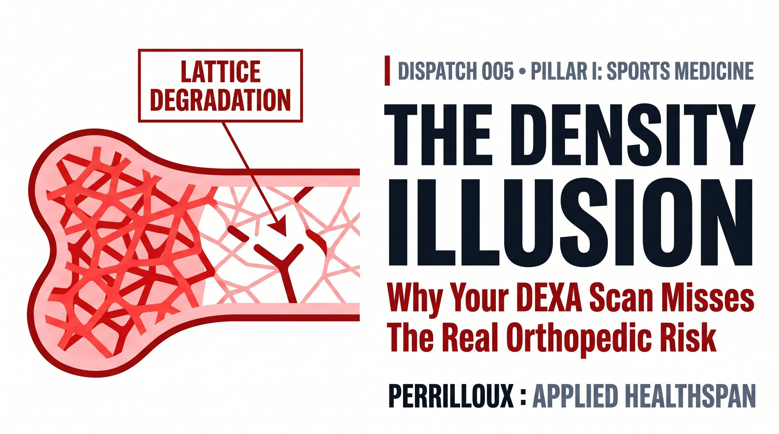
THE DENSITY ILLUSION: WHY YOUR DEXA SCAN IS HIDING YOUR TRUE ORTHOPEDIC RISK
Translating Dr. Harold Frost’s Mechanostat Theorem on Piezoelectric Mechanotransduction, Trabecular Micro-Architecture, and Osteogenic Loading for Structural Durability.
Author: Darryl M. Perrilloux, CSCS | Executive Director, Perrilloux Performance
Clinical Pillar: Pillar I: Sports Medicine & Orthopedics
Category: Applied Healthspan / Orthopedic Durability & Mechanobiology
Time: 7 Minutes
In all high-performance environments—whether designing an aerospace fuselage or optimizing a human chassis for elite athletic performance—we utilize structural telemetry as our risk-assessment metric. The notion of building a skyscraper by “estimating” load-bearing capacity is absurd, and yet, many high-performing individuals have been assessing their skeletal risk with an incomplete proxy metric: bone mineral density (BMD).
Standard annual check-ups use Dual-Energy X-ray Absorptiometry (DEXA) scans to determine the amount of pure mass in each individual's bone density. DEXA assumes that if patients maintain their bone mass at or above acceptable levels for their age group, they will be able to manage their orthopedic risk appropriately. In reality, however, annual standard physicals have serious clinical limitations.
Mechanism of Action: How Does Bone Actually Absorb Kinetic Force?
The skeletal system's ability to withstand impact without fracturing depends upon both cortical bone (the harder, denser surface layer) and trabecular bone (the inner honeycomb structure of the skeleton). The current methodologies for testing this capability — like the DEXA test — measure how much the concrete weighs. A high bone mass reading on your DEXA indicates that you have strong bones, which doesn't necessarily indicate that they will be able to resist a high-velocity impact. In actuality, whether or not an individual's bones are capable of resisting fracture in the event of an impact is determined by the geometric connectivity of the internal "rebar" (trabecular micro-architecture).
The Architectural Lattice:
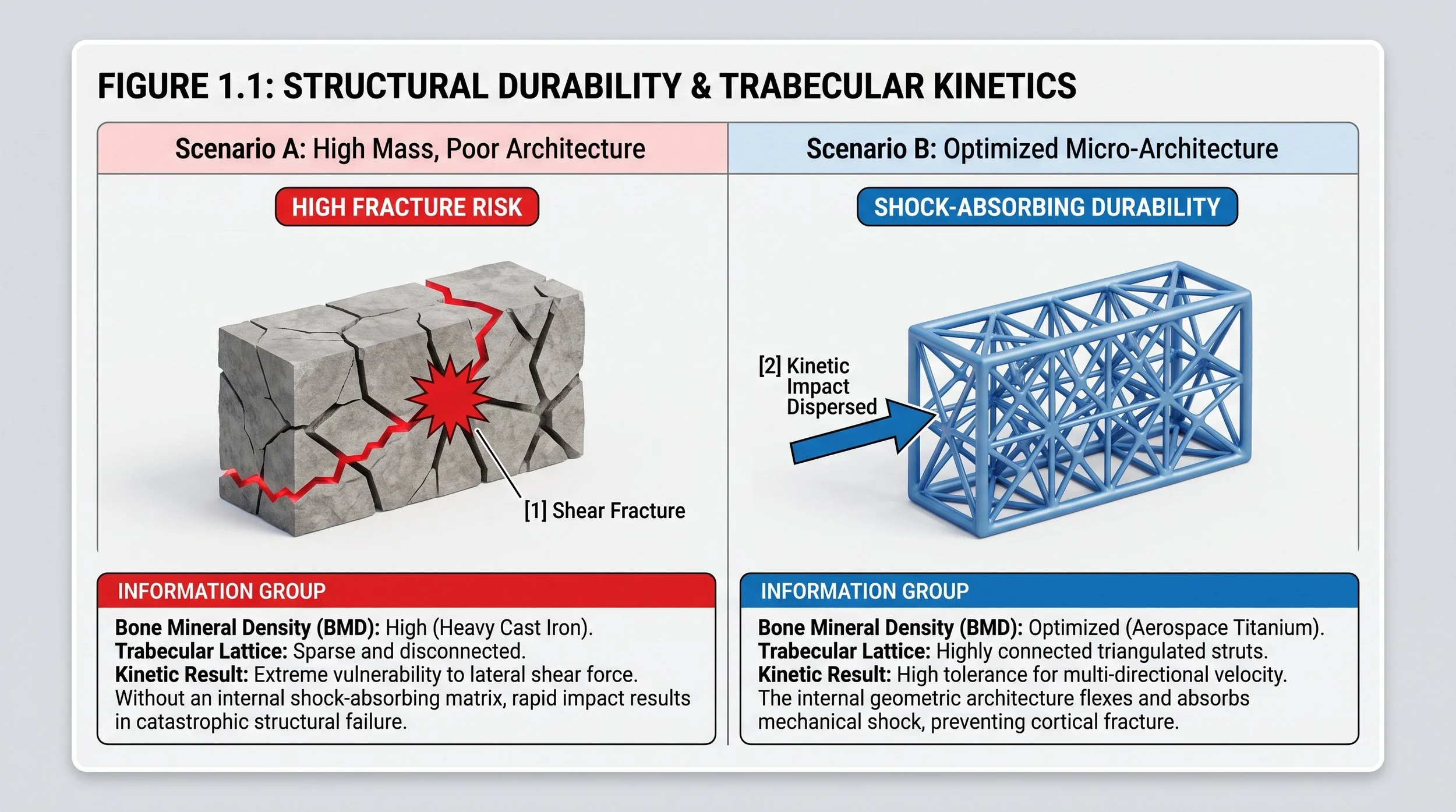
Consider your skeletal structure as an aerospace wing.
Scenario A: (High Mass, Poor Architecture). The iron wing is extremely heavy (due to high bone mineral density); however, when subjected to extreme shearing forces, it will crack and shatter.
Scenario B: (Low Mass, Optimized Architecture). You use a lightweight, 3D-printed, triangulated titanium lattice for your wing. Although this is largely composed of empty space (low bone mineral density) it can withstand massive kinetic shock because of its ability to absorb and distribute energy without breaking.
The Perrilloux Protocol: Structural Telemetry Translation
Skeletal degradation is a condition resulting from an absence of high-velocity mechanical strain over time. For all orthopedic pathology, clinical durability is determined by the total threshold of kinetic force the skeletal lattice can absorb and disperse.
Mechanistic Translation: The concept of bone remodeling should be viewed similarly to how engineers view piezoelectricity. Bones are piezoelectric materials—when they undergo rapid mechanical deformation (bending or compressing under impact), they generate an electrical charge. This electrical signal is the only language your osteoblast cells understand. Without a rapid, high-voltage mechanical spark, osteoblasts will not build new internal structural struts.
1. Laboratory Test Upgrade (The Telemetry Check):
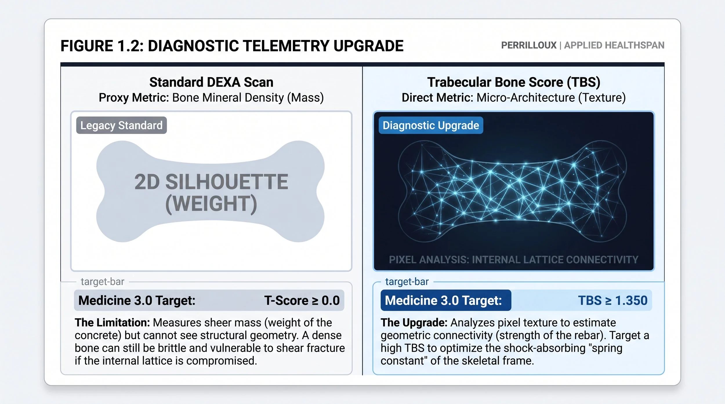
Ask your physician to include a Trabecular Bone Score (TBS) software overlay with your next DEXA scan. While DEXA measures mass, TBS evaluates the pixel texture of the scan to estimate the bone's actual microarchitectural quality. Shift from making decisions based solely on proxy weight (BMD) to understanding the geometric integrity of your skeletal lattice.
2. Baseline Calibration (The Targets):
Standard Reference: Focusing exclusively on a T-score > -1.0 (Normal Density).
Targeted Healthspan Reference: Achieving a high Trabecular Bone Score combined with functional metrics of power absorption (e.g., reactive strength index). Medicine 3.0’s target is engineered to maximize the "spring constant" of the chassis, not just its stationary weight.
3. Structural Strategy: The Pharmacokinetics of Osteogenic Loading
Bone responds to specific kinetic stimuli; however, as an osteogenically loaded stimulus, it is not intended to be a "workout" that results in excessive sweating and/or muscle fatigue. The purpose of this type of stimulus is to provide a precise mechanical dose to the skeletal frame. Therefore, applying general "fitness" principles such as "supersets", "high repetition", or "short rest period" will create excess metabolic acidity (i.e., lactate), which can interfere with the necessary neural transmission to facilitate bone remodeling. There are two major intervention points needed to promote bone growth: high-velocity impacts to initiate mechanotransduction, and Heavy Axial loads to stimulate cortical thickening.
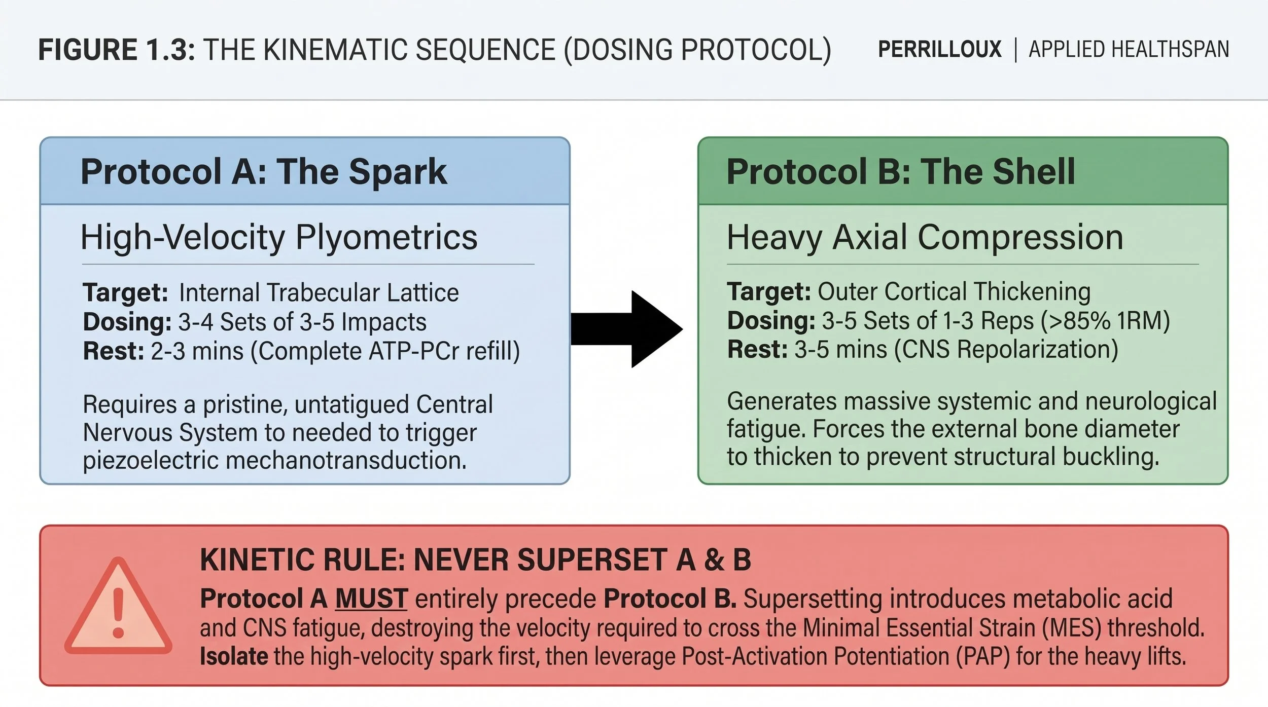
Protocol A: High-Velocity Kinetic Impact (The Piezoelectric Spark):
Biologic Mechanism: Slow, heavy lifting increases mass, but fluid shear stress inside the bone—created only by rapid acceleration and deceleration—is required to signal osteocytes. This triggers the Wnt/β-catenin signaling pathway, forcing osteoblasts to lay down new trabecular struts.
Exercise Selection: 1 to 2 movements (e.g., Depth Drops, Lateral Bounding, Max-Height Pogo Jumps).
Dosing (Sets/Reps): 3 to 4 sets of 3 to 5 impacts. The stimulus must be rapid and multi-directional. The total volume must remain strictly within 20–30 ground contacts to prioritize rate of force development rather than metabolic fatigue.
Refractory Period (Rest): 2 to 3 minutes between sets. The ATP-PCr energy system must be fully replenished to ensure that every single impact reaches the maximum Rate of Force Development (RFD) required to cross the Minimum Essential Strain (MES) threshold. If the velocity drops by even 5%, the set is terminated.
Protocol B: Heavy Axial Compression (Cortical Thickening):
Biologic Mechanism: While plyometrics build the internal lattice, subjecting the spinal column and appendicular skeleton to loads exceeding standard operational capacity forces the body to thicken the external cortical diameter to prevent structural buckling.
Exercise Selection: 1 to 2 compound axial movements (e.g., Hex-Bar Deadlifts, Heavy Yoke/Farmer's Carries, Heavy Isometric Squat Holds).
Ramp-Up Protocol (Thermal Stabilization):
Supramaximal loads can never be applied to unwarmed or "cold" soft tissues. A precise ramp-up must precede every Work-Set. The purpose of this ramp-up is to optimize both the viscosity of all soft connective tissues in your body and the slew rate of your central nervous system (CNS). Note: These are not fatiguing work-sets; they are CNS "checks."
Ramp-Up Set 1: 8 repetitions @ 50% target load (Ensure you maintain proper movement patterns throughout the set).
Ramp-Up Set 2: 5 repetitions @ 70% target load (Build force through the kinetic chain).
Ramp-Up Set 3: 2 repetitions @ 90% target load (Verify neutral).
Dosing (Sets/Reps): 3 to 5 Work-Sets of 1 to 3 repetitions, or 10-second maximal isometric holds. The weight must genuinely threaten structural capacity (> 85% of 1-Repetition Maximum).
Refractory Period (Rest): 3 to 5 minutes between Work-Sets to allow complete Central Nervous System (CNS) repolarization.
The Kinematic Sequence: Linear Execution (No Supersets)
Protocol A must be completed in full before Protocol B on the same day. Do not superset these protocols. Supersets cause systemic fatigue (signal noise). High-velocity plyometrics require a pristine Central Nervous System (CNS) to generate the peak neurological voltage needed to trigger the piezoelectric spark. Heavy deadlifts performed first or during plyometrics lead to massive CNS fatigue, destroying the velocity required to cross the MES threshold. You must isolate the high-velocity spark first. After you have neurologically primed the trabecular lattice, move to heavy axial compression. Executing them sequentially on the same day serves as Post-Activation Potentiation (PAP), in which the high-velocity jumps "wake up" the high-threshold motor units required to safely lift heavy axial loads.
Cross-Pillar Integration: The Nutritional Supply Chain
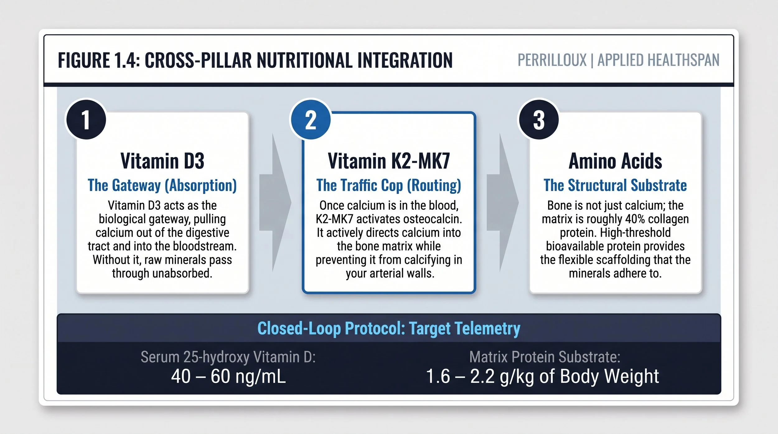
The osteogenic load of Pillar I will signal the osteoblast to begin its function as a builder; however, without providing the osteoblast with raw material from the integration of Pillar V (nutrition), it cannot perform its function. Therefore, high-velocity loading must be performed in conjunction with the appropriate systemic supply. Perrilloux Performance does not endorse a "one size fits all" or blind dosing protocols. Rather, we believe that optimization is achieved by reaching Target Serum Telemetry levels.
Closed loop supplementation protocol: test, don't guess. High velocity loading needs to be correlated with precision in Serum testing. We do not give you a dosage prescription...we give you a target state. Your physician must order blood tests to establish baseline levels before you begin taking supplements.
Vitamin D3 + K2-MK7: D3 allows for calcium absorption, while K2-MK7 is the biological traffic cop directing the calcium into the bone lattice instead of your arterial walls.
Target Telemetry (Medicine 3.0 Standard): Serum 25-hydroxy Vitamin D levels should be within 40–60 ng/ml. Increase dose according to baseline telemetry until the desired range is achieved.
Bioavailable Amino Acids: The bone matrix contains approximately 40% collagen protein. Daily protein thresholds (target: 1.6 g/kg to 2.2 g/kg of body weight) will provide the matrix substrate necessary for structural remodeling.
The Frontier: Active Clinical Research
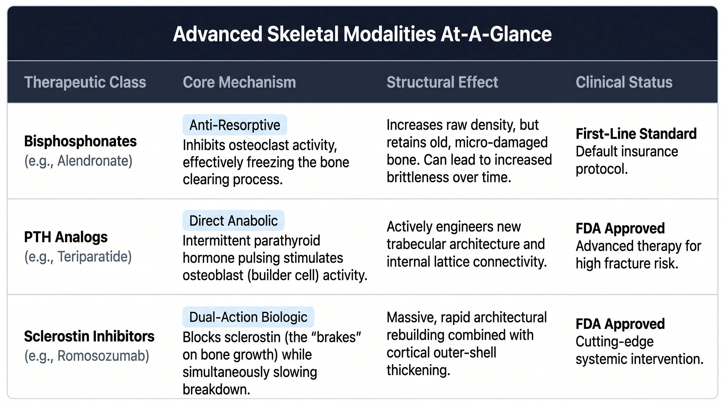
The biopharma landscape for skeletal preservation has shifted from simply freezing bone loss to actively engineering new bone.
Anti-Resorptive Agents (Bisphosphonates): Medications like Fosamax act by inhibiting osteoclasts (the cells that resorb old bone). While this keeps density high on a DEXA scan, it leaves old, brittle, micro-damaged bone in the skeleton. It increases mass but degrades architectural flexibility.
Anabolic Agents (PTH Analogs and Sclerostin Inhibitors): Unlike bisphosphonates, they promote new bone formation. The drug teriparatide (Fortéo) stimulates osteoblasts through pulsatile release of parathyroid hormone, whereas romosozumab (Evenity) has two effects. It is both a PTH analog and a sclerostin inhibitor. Sclerostin acts like the brakes on bone growth, so when it is inhibited, the result is an increase in both the formation of new trabecular architecture and a decrease in the rate at which existing bone is broken down.
Request that your physician order a Trabecular Bone Score (TBS) software overlay to be used with your next DEXA scan. Unlike BMD, which measures the density (mass), TBS uses pixel analysis to assess the actual micro-architecture (texture) of your bones. Begin to transition away from using proxy weight (BMD) as the sole basis for decision-making in relation to your skeletal integrity.
APPENDIX: THE CLINICAL EVIDENCE BASE
In this issue, we take peer-reviewed mechanobiology and translate it into applied structural protocols.
Frost, H.M. (2003). "Bone's mechanostat: a 2003 update."
Clinical Data: Frost’s theory demonstrated that bone structure is controlled by mechanical strain thresholds. When strains are less than the minimum essential strain (microstrain µε), bone will resorb. Conversely, when mechanical strains exceed the minimum essential strain, bone structure is modeled.
Translation Architecture: At Perrilloux Performance, we help our clients develop a plan to avoid “maintenance” type exercises that cause minimal impact. Instead, we create a pre-active loading model for our client that exceeds the mechanical threshold with specific plyometric and axial loading interventions that stimulate biological structural adaptation.
The Bottom Line for High-Performing Professionals & Elite Athletes
Orthopedic durability establishes the structural foundation that will support elite levels of bio-energetic performance.
The Professional: Physical fragility creates a significant obstacle for long-term health. A single accident resulting in a fractured pelvis/femur is all it takes to initiate an acute onset of immobility, metabolic failure, and cognitive degradation. Constructing a bone lattice with sufficient shock-absorbing capacity guarantees mobility well beyond age 90.
Elite Athlete: The ability to produce immense horsepower via muscle contraction is irrelevant unless the athlete has an appropriate skeleton to accommodate such large amounts of force/torque generated by the muscles. Stress fractures and tendon ruptures can result when power output exceeds the skeletal structure's ability to withstand the resulting forces. Proper osteogenesis (formation of new bone) through precise load-bearing on the bones will ensure the bone lattice remains structurally intact while operating at high RPMs (red line).
PERRILLOUX PERFORMANCE: CLINICAL ARCHITECTURE
This dispatch represents an active operational module within the Five Core Pillars of the Perrilloux Performance clinical architecture.
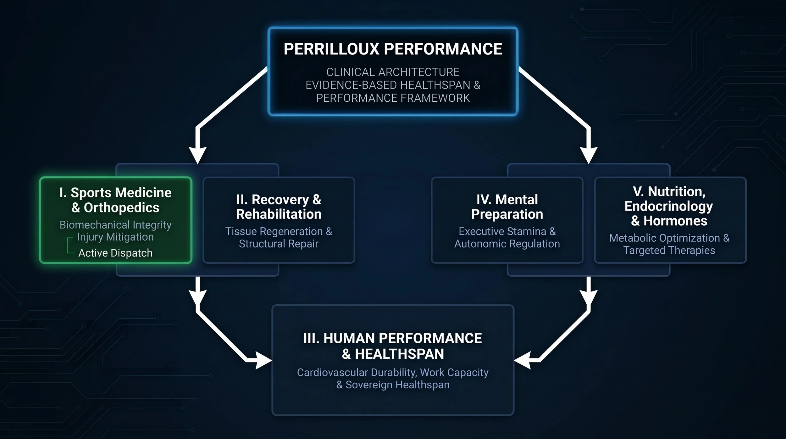
[X] I. Sports Medicine & Orthopedics (Biomechanical integrity & injury mitigation)
[ ] II. Recovery & Rehabilitation (Tissue regeneration & structural repair)
[ ] III. Human Performance & Healthspan (Cardiovascular durability & bioenergetics)
[ ] IV. Mental Preparation (Executive cognitive stamina & autonomic regulation)
[ ] V. Nutrition, Endocrinology & Hormones (Metabolic optimization & targeted therapies)